
Danish Researchers develop Novel Method for Stroke Assessment
A sophisticated scanner has provided a detailed image of the biochemistry underlying stroke. And this could be extremely significant in ensuring the best possible treatment for stroke patients.
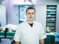
Professor Christoffer Laustsen, Head of the MR Research Centre at the Department of Clinical Medicine, Aarhus University.
It is important to begin treatment as quickly as possible when someone suffers a stroke – in other words, a clot or haemorrhage in the brain.
The faster we can stop the bleeding or remove the clot, the better the chance of limiting – and in the best case, avoiding altogether – permanent impairment in the stroke patient.
It is a well-known fact that impairment can occur when the supply of blood and oxygen to parts of the brain is reduced or constricted after a stroke.
However, when it comes to the so-called time window after stroke onset, there are many unanswered questions. This window is the time within which doctors must begin treatment if there is to be any hope of helping the patient and preventing serious side effects.
As yet, there is no clear answer as to how long this window lasts. A rule of thumb is that the window is open for a few hours after stroke onset. However, this is by no means a precise time frame, and it does not take into account individual vulnerabilities related to brain damage caused by a stroke.
If we can gain a better understanding of these vulnerabilities, we will eventually be able to assess whether a stroke patient could benefit from treatment – even if treatment is not started until after “the first few hours”.
‘This will enable us to provide more personalised stroke treatment than is possible today, and it could extend the time window for some patients,’ says Professor Christoffer Laustsen, Head of the MR Research Centre at the Department of Clinical Medicine, Aarhus University.
And there is much to indicate that this will be possible, as demonstrated in animal trials – with pigs – conducted by Professor Laustsen and his colleague Nikolaj Bøgh, doctor and Ph.D. student. Their work was recently published in the scientific Journal of Cerebral Blood Flow & Metabolism.
Several other scientists from the MR Research Centre at Aarhus University and researchers from the Technical University of Denmark and the University of California, San Francisco, were also involved in the study, which received funding from the Lundbeck Foundation.
A Rare Scanner
In 2018, Professor Laustsen was awarded a Lundbeck Foundation Fellowship worth DKK 10 million to study the basic mechanisms of brain cell metabolism.
These are biochemical mechanisms – alterations in brain cell metabolism – and are a kind of “metabolic fingerprint”.
To measure these, Professor Laustsen and Nikolaj Bøgh – who was the lead author of the scientific article – used hyperpolarised magnetic resonance imaging (MRI).
“We needed a special scanner to do the job, and there are only 24 of these in the world. Two happen to be in Denmark: one at Rigshospitalet, University of Copenhagen, and one here at Aarhus University,’ Nikolaj Bøgh explains.
Like other medical scanners, these MRI scanners provide three-dimensional images of the subject under examination. However, hyperpolarised MRI produces additional information, such as where the biochemical substances are going and what they are converted to.
‘This creates the metabolic fingerprint that is key to further development of the method for stroke assessment demonstrated in our scientific article,’ says Professor Laustsen.
In the animal trial, on which the article is based, the researchers injected ten pigs with a drug to provoke stroke.
Christoffer Laustsen and Nikolaj Bøgh then used the sophisticated scanner to measure a long list of metabolic factors in the pigs’ brains, and they repeated the measurements during the subsequent hours.
‘The metabolic fingerprints we were able to draw based on all these scans tell an extremely interesting story,’ says Nikolaj Bøgh:
‘We were able to divide the types of impairment suffered by the pigs after the stroke into several categories. And we could prove that one category of impairment will show positive development over time – in other words, the impairment will decline. In the case of another category, the impairment deteriorates.’
But how realistic is it to assume that there will be the same reactions to stroke in humans?
Professor Laustsen believes that there is a high likelihood of this:
‘We used hyperpolarised MR imaging to measure healthy people, and the results were basically the same as for the healthy part of the brains of the lab pigs.
We will now ask a number of patients admitted to hospital with a stroke for their permission to perform a scan. We’ll then see whether pigs and humans also react similarly in this situation,’ says Professor Laustsen.